COVID-19 Autopsies
Vivek Sekhawat
Sebastian B Lucas
The ongoing global COVID-19 pandemic has brought into the spotlight the critical role and necessity of autopsy. The rapid spread of infection has resulted in overload of healthcare services internationally, and the medical community continues to face difficulties in developing effective treatments for a disease pathophysiology that is incompletely understood. Several factors have impacted the ability of pathologists to respond to this need. Understandable concerns for safety and limiting the spread of infection, particularly early on in the crisis when transmission was less fully understood, have meant that fewer autopsies could be justifiably performed, within both the Coronial and hospital settings in the UK. In addition, the burdens on healthcare services have imposed significant resource limitations. Mortuary services have been under tremendous strain in managing the flow of bodies, and clinical teams have worked in trying conditions with restrictions placed on visiting relatives, which has affected communications and the ability to acquire consent for autopsies.
I (VS), as a histopathology registrar, have been working within a team of two autopsy pathology consultants and APTs performing hospital autopsies on a limited number of COVID-19 positive patients since late April, with several objectives – foremost, to try to elucidate for clinicians the pathophysiology leading to death in their individual patients, and further our understanding of this disease. We have also been working with clinical colleagues in service improvement projects, acquiring samples of synovial fluid and ophthalmic fluids for virological analysis to inform safe planning of the resumption of orthopaedic and ophthalmology services. Finally, colleagues in translational research settings are heavily reliant on pathologists in the consented acquisition of tissue, for their work which may ultimately help develop effective treatments, tests and vaccines. 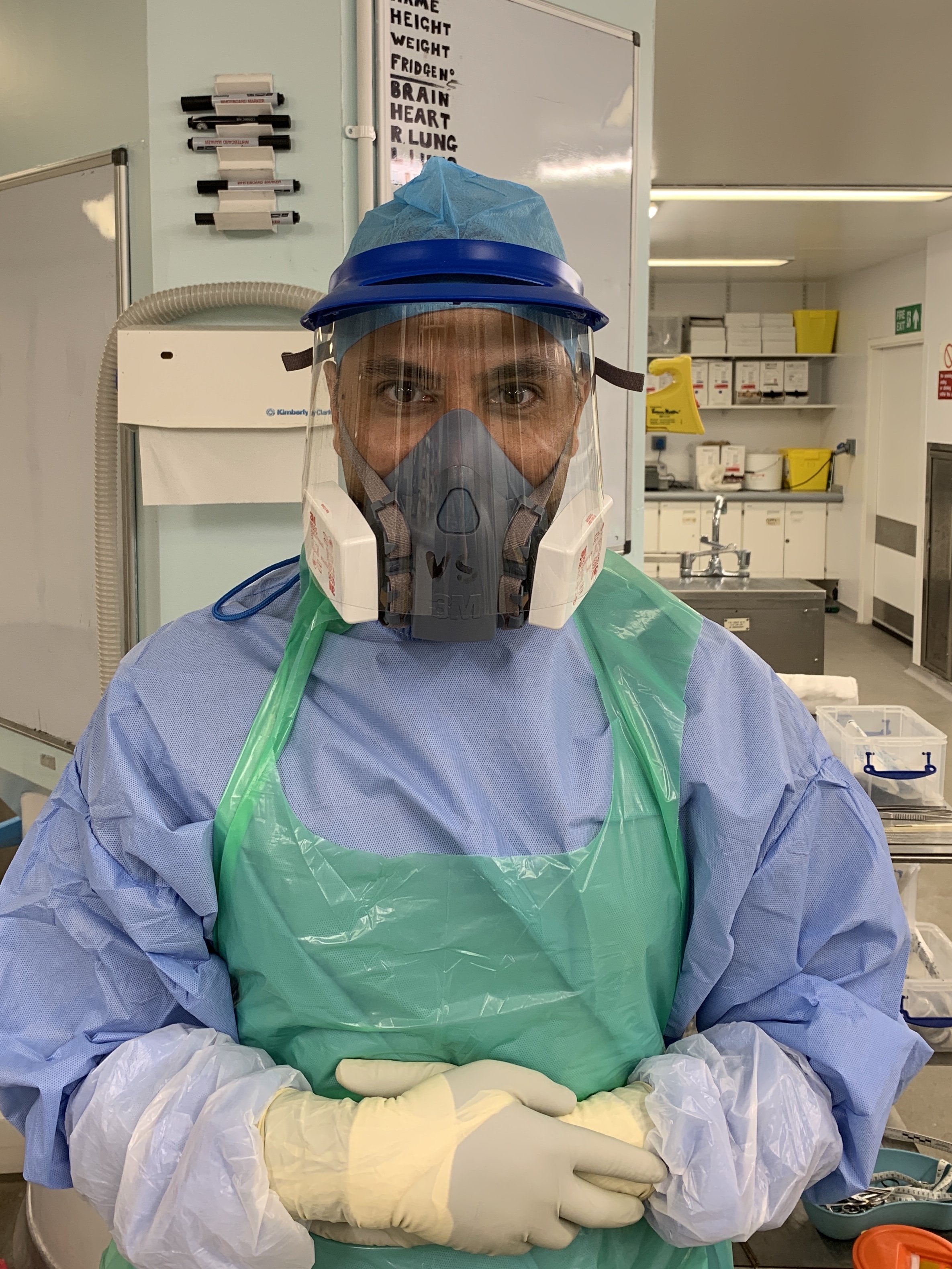
This has required the development of an extensive procedural and sampling protocol, and working under full PPE has been at times physically exhausting and left us with sore nasal bridges! In order to maximise efficiency and minimise the duration of exposure of the whole team, I have taken the role of clean circulator within the mortuary, assisting in sampling, documentation and photography. As a trainee, being involved in all steps of the process, from the remote consenting process, to development of the sampling protocols, the specialised autopsy procedure, and histological assessment and working up the cases, has been an invaluable learning experience.
The published autopsy literature in COVID-19 positive patients to date has revealed some common pathological themes, and general overview now follows.
Macroscopic venous thrombosis has been reported in several patients, in line with clinical reports of increased incidence of thrombo-embolic events in COVID-19 patients.
The lungs are heavy, firm and congested, though in earlier stages may show relatively normal weights. Histologically there is diffuse alveolar damage in varying phases of evolution. Early cases may show widening of alveolar septa, capillary and vascular congestion, mild interstitial mononuclear inflammatory infiltrates, increased circulating megakaryocytes, and platelet-fibrin microthrombi. There is progression with hyperplasia and desquamation of type 2 pneumocytes with atypia (but which have not so far shown convincing viral inclusions) and increased alveolar macrophages, both of which have been demonstrated in several studies to contain SARS-CoV-2 viral RNA and antigens. Associated hyaline membrane formation and varying degrees of organisation and fibrosis appear to correlate with the length of disease course, with more advanced features in hospitalised patients. Squamous metaplasia is variably observed. Large vessel thrombosis/thrombo-embolism is also seen, sometimes with parenchymal haemorrhagic infarction. Superimposed bacterial pneumonia is reported. Invasive aspergillosis may complicate pneumonitis in the ITU setting, and such infections have been previously associated with ECMO therapy.
Hearts generally show non-specific features of pre-existing comorbidities, such as hypertension, ischaemic heart disease and diabetes. Intramyocardial microthrombi may be seen. Occasional individual myocyte necrosis is seen along with interstitial oedema, and whilst sparse interstitial mononuclear inflammatory cell infiltrates have been observed in association, convincing evidence of myocarditis has not been seen in the majority of published series. Direct viral infection of myocytes has also not been demonstrated.
The haematolymphoid system shows T-cell depletion and a reactive lymphoplasmacytosis with variably atypical/activated morphology. Occasional parenchymal necrosis of lymph nodes is seen. There is white pulp atrophy and red pulp expansion within the spleen, sometimes associated with haemophagocytosis. Lymphoid depletion generally appears less pronounced than was seen in SARS (SARS-CoV, 2003). Bone marrow may show left-shifted granulopoiesis and haemophagocytosis.
Kidneys show features of acute tubular injury in most cases with vacuolation of proximal tubular epithelium, and intracytoplasmic viral particles have been demonstrated by electron microscopy. Viral particles have also been seen within podocytes. Pigmented myoglobin casts may be present. Glomerular changes appear to generally correlate with pre-existing disease, and glomerular microthrombi have been identified in only a minority of cases, whereas microthrombi in vasa recta and medullary vasculature are sometimes present.
The liver shows features of passive congestion and may show Kupffer cell activation and secondary haemophagocytosis.
The central nervous system has been examined less widely in autopsy series, but some have reported minimal neuronal necrosis, hypoxic injury and vascular and demyelinating lesions. No direct evidence of viral encephalitis or meningitis has been reported. Viral particles have however been reportedly observed by electron microscopy in neural cell bodies and cerebral endothelial cells.
Other organs predominantly show shock sequelae.
Some groups have described features of microvascular and endothelial injury, in several organ tissues. Vasculitis however has not been generally observed.
There is currently much ongoing research into the procoagulant state that seems to occur in at least a subset of these patients. Hypothesized aetiologies include non-specific endothelial injury as seen in other respiratory infections with associated pulmonary tissue factor release, sepsis and cytokine storm, as well as more specific interactions between virally-infected cells, the renin-angiotensin and complement systems, and even neutrophil extracellular traps. Whilst clinical studies have reported increased incidences of disseminated intravascular coagulation defined by clinical parameters, there does not appear to be much pathological evidence to support a widely disseminated “consumptive” coagulopathy, particularly given the infrequent glomerular involvement. Microthrombosis appears to be more prominent in specific organs, notably within the lungs and heart; whether this involves the central nervous system remains to be seen. It is also noted that glomerular endothelium, unlike other systemic endothelia, has been shown to have reduced expression of the ACE2 receptor, by which SARS-CoV-2 (and SARS-CoV, 2003) gains cellular entry, which could provide a potential explanation for a more tissue-specific thrombotic microangiopathy.
Mortuary teams have worked tirelessly to support this work around the unprecedented logistical challenges they have faced during the pandemic. We have found relatives of deceased patients to be extremely generous, and overwhelmingly express a desire to help their fellow citizens by increasing scientific understanding, despite very obviously difficult circumstances. The emergency legislative changes and adaptations within the coronial system, with no immediate end to the crisis within sight, raise concerns about the future of autopsy practice within the UK. Now is therefore a critical time for the pathology community to demonstrate the essential value of autopsy.